


Squamous cell carcinoma (SCC) is considered one of the most prevalent malignant tumors in the skin as well as in areas with mucosal surfaces. It develops from the squamous epithelial cells and is marked by incessant proliferation and a tendency to infiltrate deeper tissues.
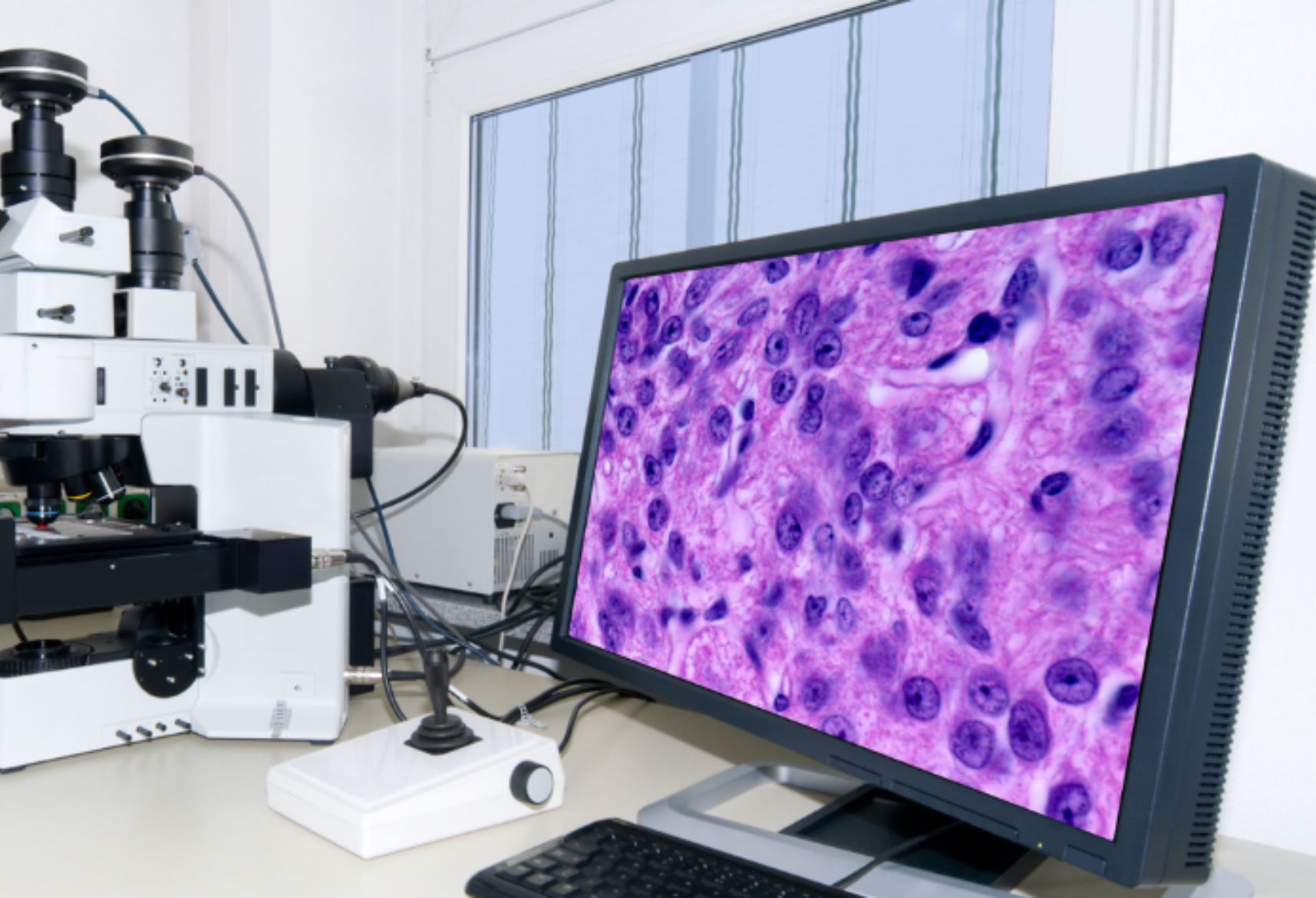
The histopathological evaluation is important in the diagnosis of SCC, its grade classification, and treatment provisions.
Histopathological Features
In the case of squamous cell carcinoma, some of these features are specific and very important from the viewpoint of diagnosis.
These include the following primary features observed in the microscope:
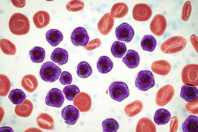
Atypical Squamous Cells
- The abnormality of squamous epithelial cells is a unique feature of SCC.
- Squamous cell carcinoma is accompanied by abnormal squamous epithelial cells which tend to have enlarged hyperchromatic nuclei and irregular nuclear membranes.
- Distinctive features include increased axially rotated nuclear within a cell cytoplasm.
Keratinization
- The presence of keratinized SCC cells with keratinized pearls or single keratinized cells indicates the possibility of keratinization.
- This feature is more pronounced in well-differentiated SCC.
Infiltrative Growth Pattern
- SCC has an infiltrative form of pancreatic carcinoma and is clinically more pronounced than benign lesions.
- Basement membrane penetration by tumor cells and subsequent invasion into the underlying stroma occurs.
Desmoplastic Stromal Reaction
- Interstitial or the surrounding stroma showing the increased activities of fibroblast and deposition of collagen is common.
- This reaction helps the tumor firm and invasive type.
Inflammatory Response
- SCC can have basal cell carcinomas with a lymphoid infiltrate rich in T and B lymphocytes.
- The immune cells are likely to be due to active tumor proliferation.
Vascular and Perineural Invasion
- Advance stages of SCC may demonstrate invasion to blood vessels and peripheral nerves.
- Perineural invasion is considered a more malignant feature and is perhaps associated with greater chances of recurrence.
Grading of Squamous Cell Carcinoma
- SCC is graded according to degree of differentiation, which has implications on treatment options and prognosis:
- Well Differentiated SCC: Contains prominent keratinized squamous epithelium and is similar to the normal one.
- Moderately Differentiated SCC: Intermediate between well and poorly differentiated SCC with keratinization.
- Poorly Differentiated SCC: Non-keratinizing, marked pleomorphism, and heightened mitotic activity is observed.
Diagnostic Techniques
- Hematoxylin and Eosin (H&E) Staining: This is the most common technique employed in assessing SCC histopathology.
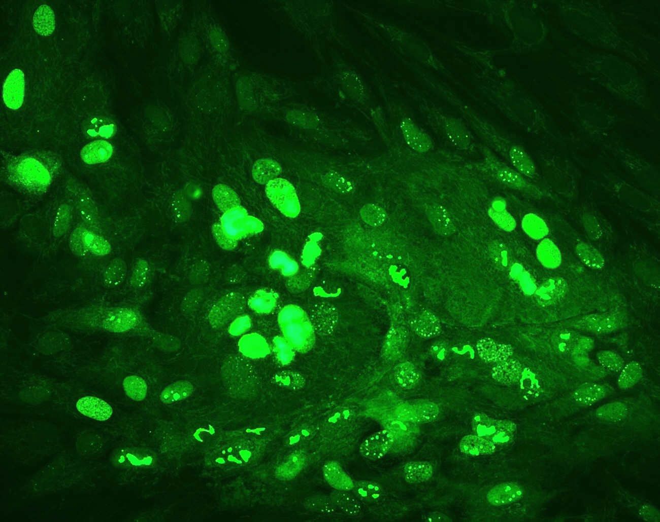
- Immunohistochemistry (IHC): p63, CK5/6, and EMA positive markers are useful in the diagnosis of SCC.
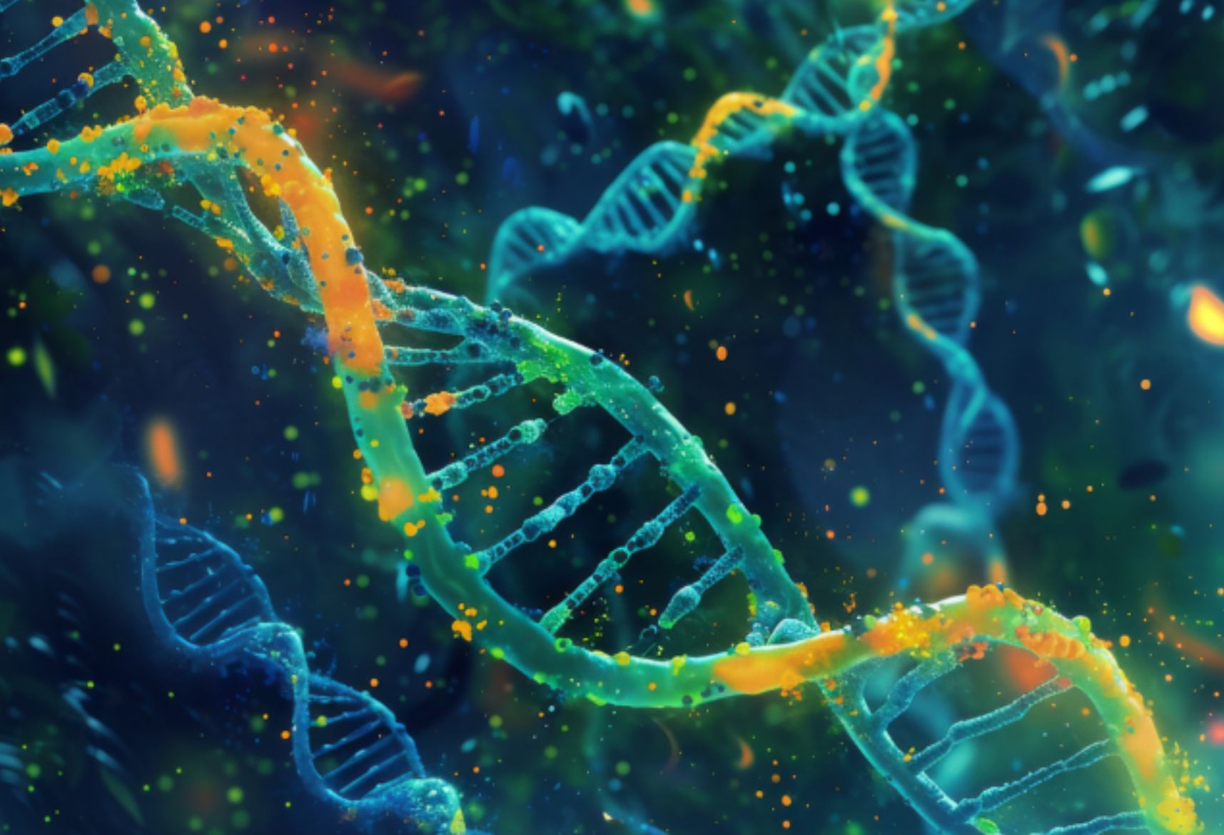
- Molecular Testing: Performed for intervention on prognosis and for targeted therapy.
Conclusion
Histopathological examination is critical in squamous cell carcinoma diagnosis, determining the grade, and making treatment options. Once effective histologic features are put into consideration, pathologist determine the aggressiveness of a tumor and the survival results of a patient in need of immediate care.