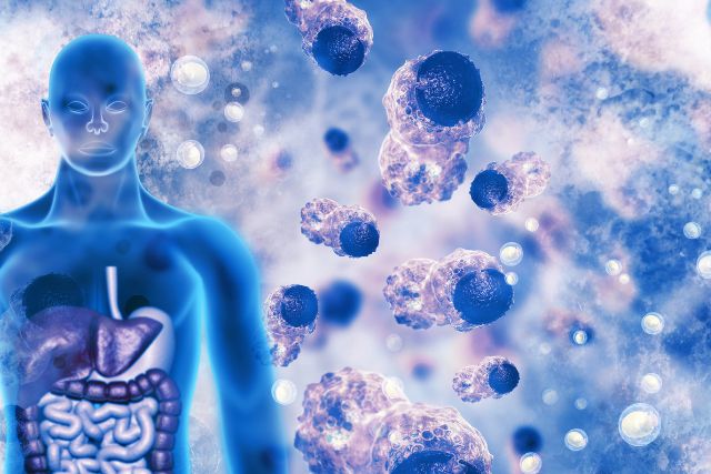


The preparation and analysis of tissue samples require the use of the cryostat, which is an essential tool in the discipline of histopathology. Histopathology is the study of diverse cellular structures as well as the microscopic inspection of tissue samples to determine the level of tissue damage and identify diseases.
Here’s a breakdown of the role of the cryostat in histopathology, from sample collection to analysis:
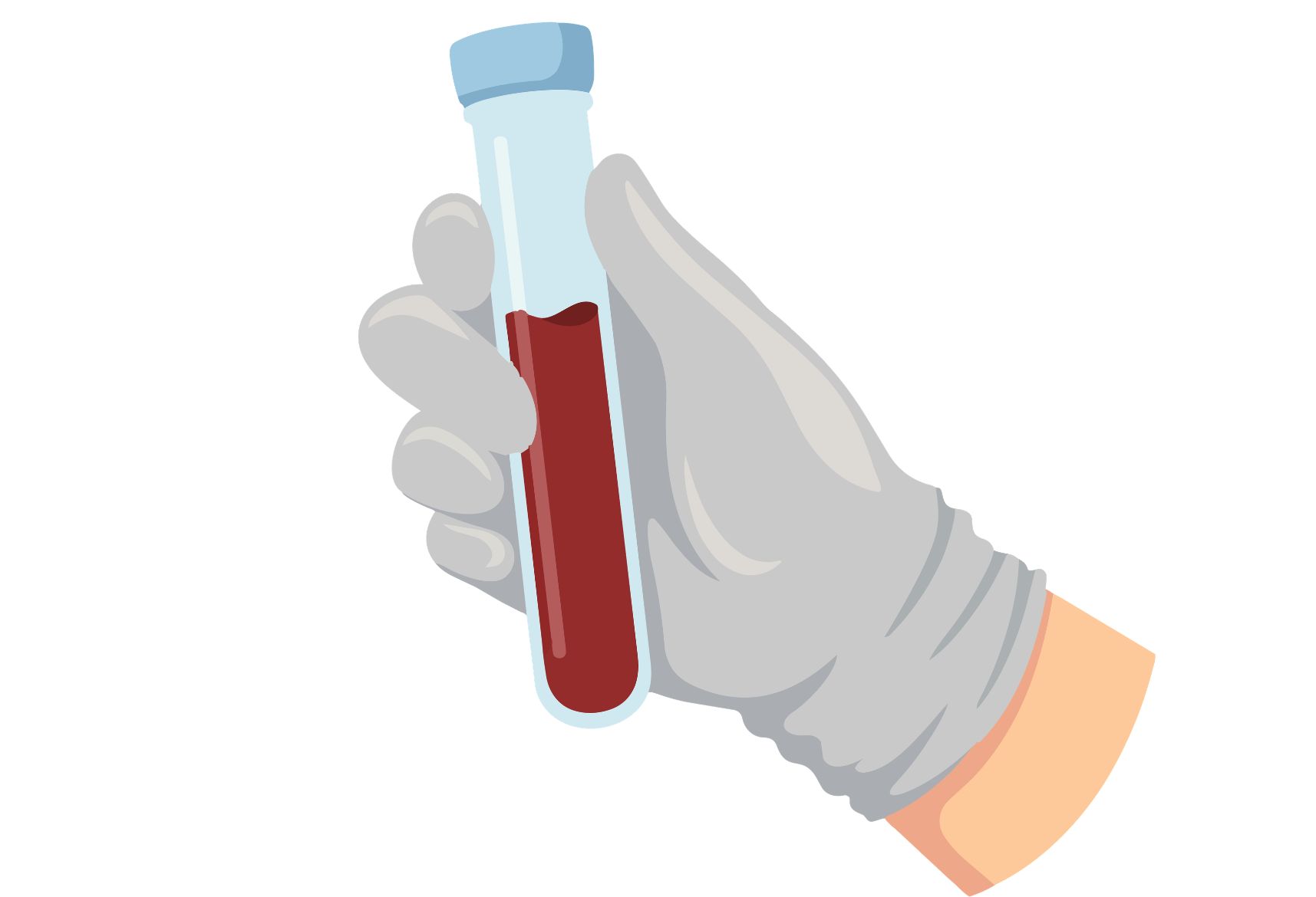
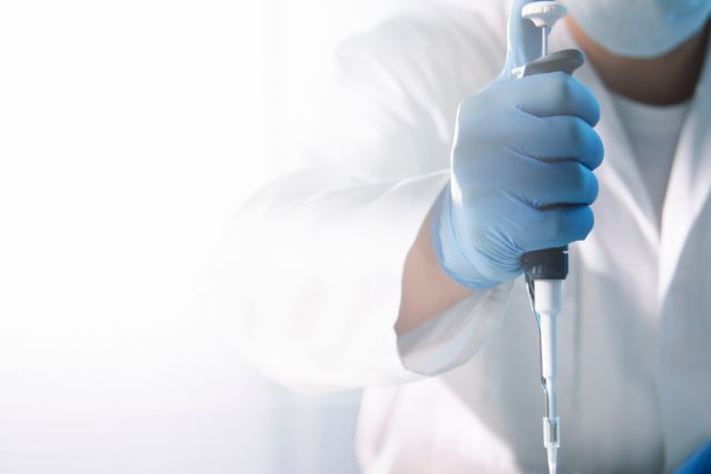
Sample Collection:
- The collecting of tissue samples, often obtained by procedures like biopsies or surgical excisions, is the first step in the histopathology process.
- Depending on the specific diagnostic or research requirements, these tissue samples may be taken from different bodily areas.
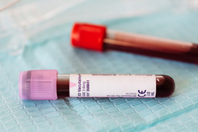
Tissue Fixation:
To retain their cellular structure and stop degradation, tissue samples are typically immersed in a fixative solution, such as formalin, after collection.
Tissue Processing:
The processed fixed tissue samples go through a number of stages, including as cleaning, dehydrating, and embedding in paraffin wax. These procedures get the tissue ready for cutting and analysis.
Sectioning:
- The tissue becomes rigid and suitable for sectioning after being embedded in paraffin wax.
- The cryostat is used to cut thin tissue sections (usually 4-7 micrometers thick) from the paraffin-embedded tissue block. These sections are sometimes known as “histological slides” or “tissue sections.”
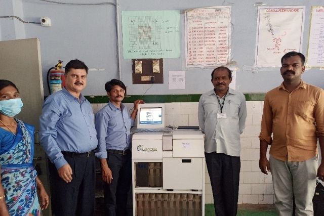
Cryostat Functionality:
- A cryostat is a type of freezing microtome that is used to segment frozen tissue specimens.
- It keeps the tissue block at a controlled low temperature (usually between -20 and -30 degrees Celsius) to harden the tissue and allow for thin sectioning.
- Within the cryostat, the tissue block is positioned on a microtome stage.
- A blade or knife installed on the microtome stage cuts thin portions from the tissue block as it is progressively advanced.
- After collecting the sections on glass slides, they are available for further processing and staining.
Staining:
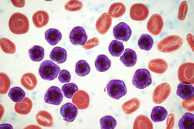
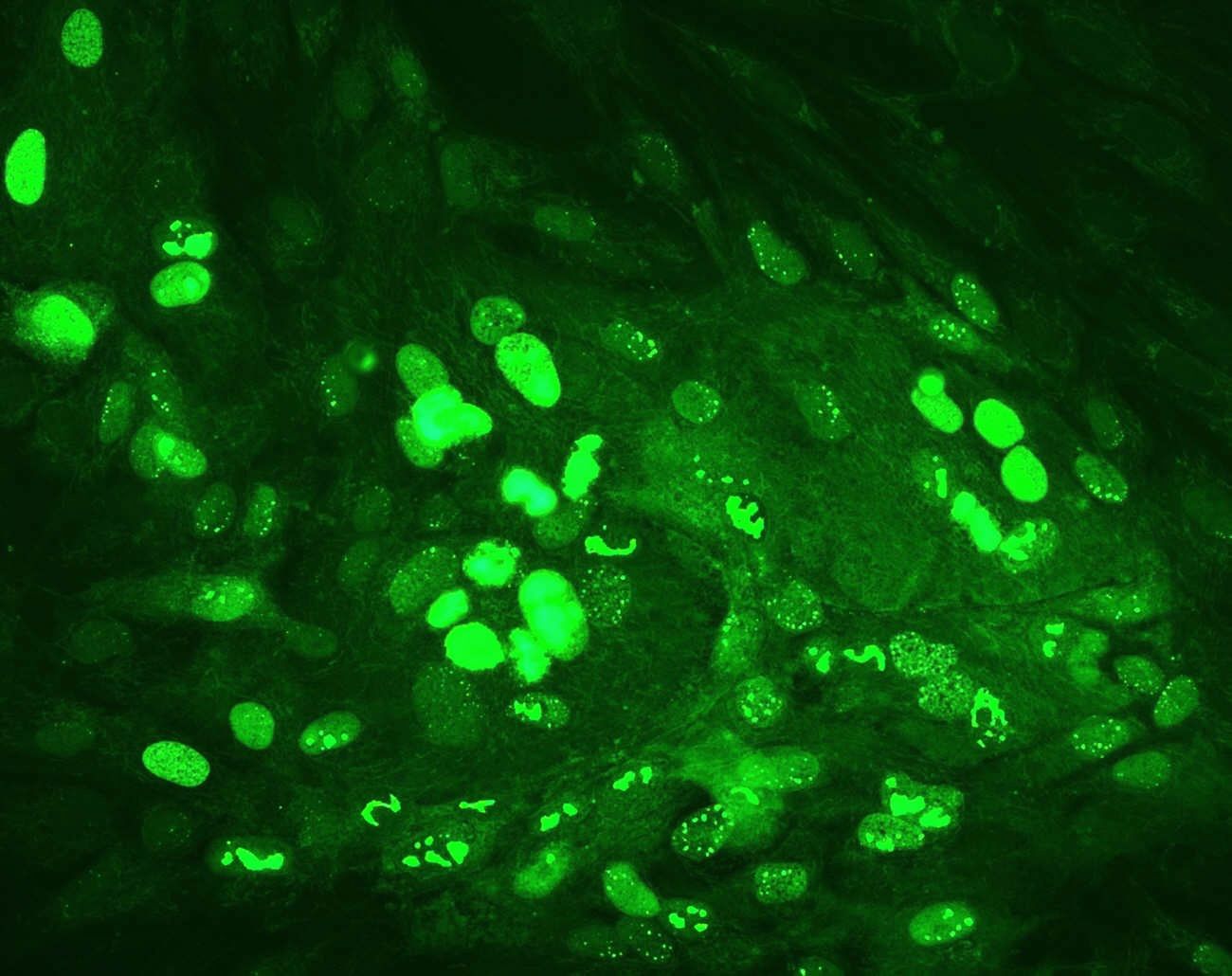
- To highlight specific cellular structures or abnormalities, tissue sections are stained with various histological stains such as hematoxylin and eosin (H&E) or unique stains.
- Staining improves the contrast of tissue sections, allowing pathologists to recognize and assess cellular characteristics more easily.
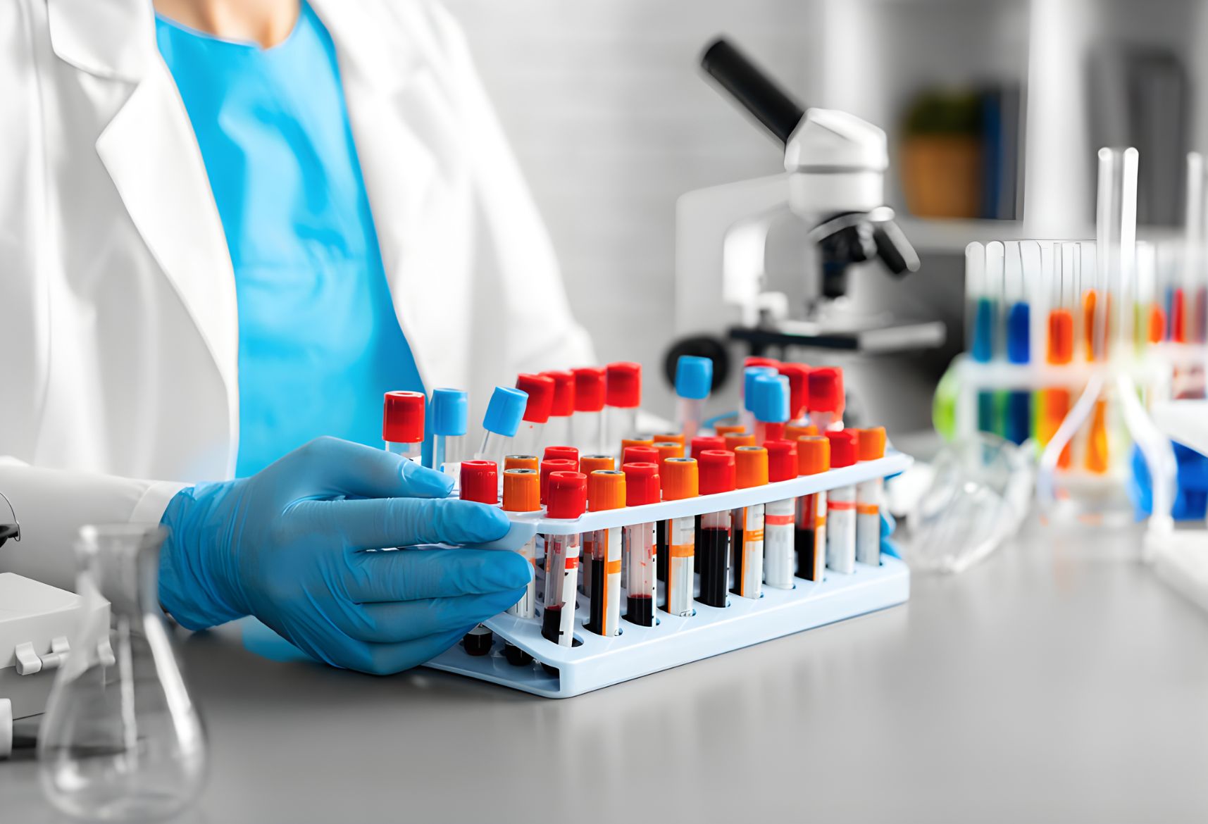
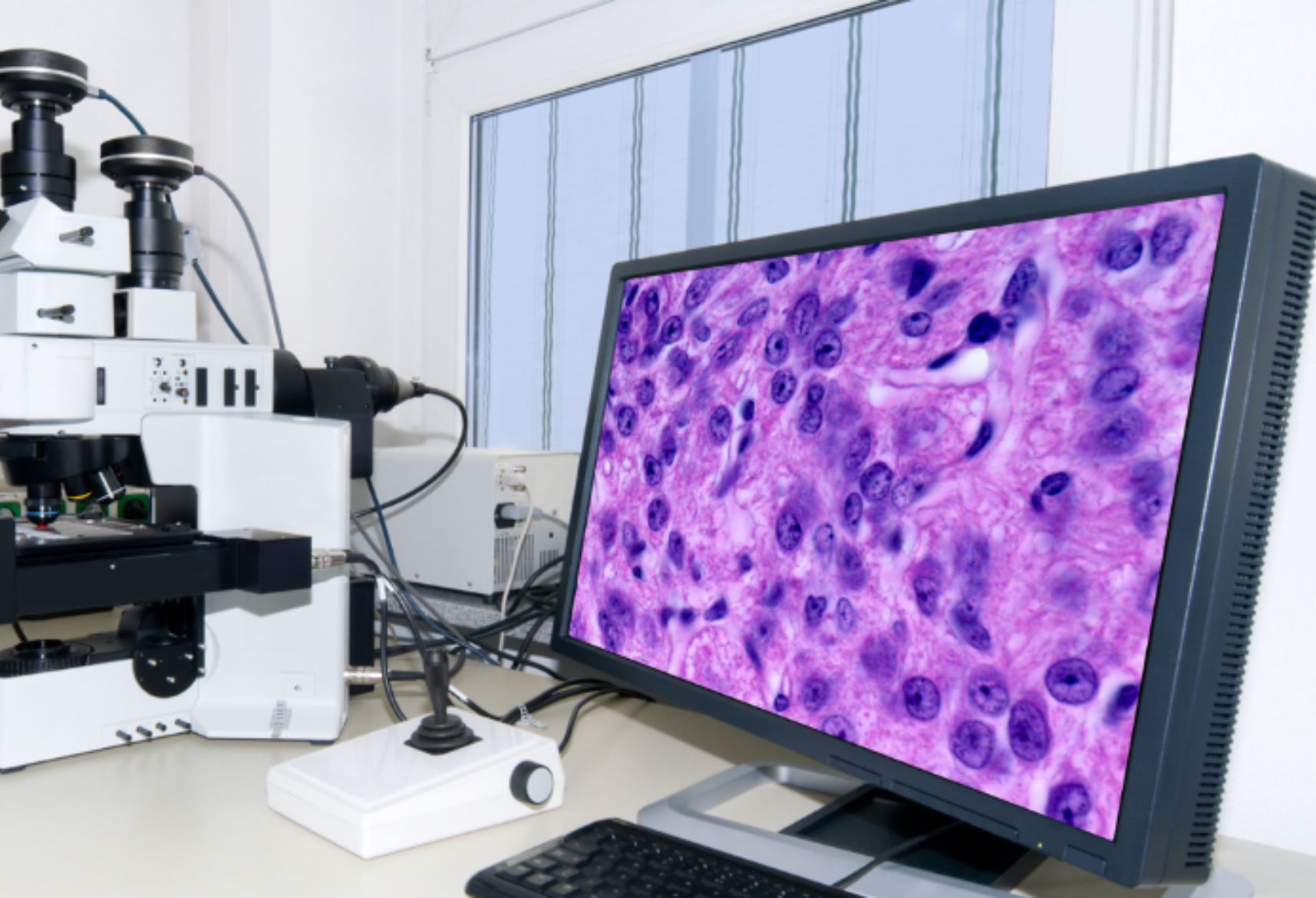
Microscopic Analysis:
- A pathologist or histotechnologist examines the stained tissue sections under a microscope after staining.
- Microscopic examination enables the evaluation of cellular morphology, tissue architecture, and the diagnosis of anomalies or illnesses.
Diagnosis and Reporting:
- The pathologist delivers a diagnosis or clinical interpretation of the tissue sample based on microscopic analysis.
- A pathology report is created, which provides facts about the diagnosis as well as disease stage, grade, and prognosis.
In summary, the cryostat plays a vital role in histopathology by facilitating the preparation of thin tissue sections from frozen tissue specimens. These sections are subsequently stained and analyzed under a microscope to diagnose diseases, evaluate tissue abnormalities, and guide clinical management. The accuracy and quality of tissue sections produced by the cryostat are essential for the reliable diagnosis and treatment of patients.