


Immunofluorescence microscopy (fluorescent antibody) was reported by Coons, Creech and Jones in 1941. In 1942, Coons and Kaplan reported that fluorescence dyes can be conjugated with antibodies. These labelled antibodies further used as probes to detect and locate antigen specific to this antibody. Coons and Kalpan in 1950 used Fluorescence microscopy for the first time. Riggs et al (1958) observed that more stable Fluorochrome was fluorescein isothiocyanate (FITC). In 1958, Goldwasser and Kissling used Fluorescence microscopy for the diagnosis of Rabies.
Definition
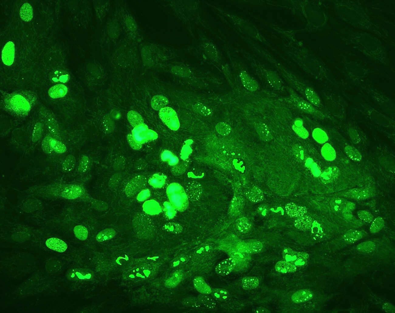
When immunofluorescence is specifically used to detect an antigen by conjugating the fluorescent dye with the Fc region of the specific antibody (immunoglobulins) against that antigen, it is referred to as Fluorescent Antibody Test (FAT).
For full text article : Click here